
Vitamin A signals and thyroid hormones reroute cone cells, reshaping birth-vision biology
A new study finds blue cones do not simply migrate, they transform into red and green under vitamin A-related cues.
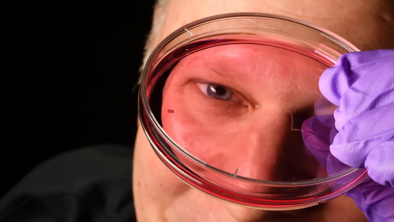
Scientists have uncovered how human sharp central vision before birth may form: blue cone cells transform into red and green cones. The discovery points to better lab-grown retinal tissue and potential cell therapies for vision loss.
Vision research has been chasing a simple question for years: how do humans build crisp central vision in the womb? A study reported through ScienceDaily changes the answer in a way that matters for both basic biology and the future of retinal repair. Instead of blue cone cells migrating away from the retina's center, the study found those cells can transform into red and green cones, guided by vitamin A-related signals and thyroid hormones. That single pivot rewires how scientists think about what is happening during development, and it directly shapes what they might be able to engineer later.
Why this matters right now is that the retina is not just an organ. It is the input layer for reading, faces, driving, and independence. If you are trying to grow or replace retinal cells in the lab, or to design cell therapies for conditions that damage vision later in life, the developmental “instruction set” is the blueprint. The study’s finding suggests the blueprint is not only about where cells go, but what they become. Vitamin A-related signaling and thyroid hormones appear to influence cone identity, turning what would otherwise be blue cone fate into red and green cone fate, specifically in the context of central vision formation.
To understand why executives and boards should pay attention, zoom out to how retinal technology usually works. Most approaches aiming to restore or preserve vision, whether surgical, pharmacological, or cellular, depend on making the right cell types at the right time and in the right microenvironment. In the past, researchers often focused on structural outcomes, like getting cells to occupy the correct retinal locations. But cell identity is its own bottleneck. If blue cones in the developing retina can transform into red and green cones instead of moving away, then the “center-building” mechanism is partly biochemical. That means lab-grown tissue might need vitamin A-related cues and thyroid hormone signals, not just generic differentiation protocols.
This is where regulation and translation logic come in. Vision therapies sit in a high-scrutiny space because they involve delicate tissue, long-term functional outcomes, and in many cases irreversible changes once cells are implanted. Regulators typically want evidence that a therapy not only survives, but behaves predictably. A development-driven mechanism like this, grounded in specific signals such as vitamin A-related signals and thyroid hormones, can help researchers define what success looks like beyond survival. If the goal is functional central vision support, the field will likely focus on whether induced or transplanted cells express the correct cone types and integrate into the retinal circuitry in a developmentally consistent way.
There is also a practical industry angle: manufacturing and differentiation processes. Lab-grown retinal tissue is hard because retina development is complex, and small deviations can produce the wrong cell mix. When a discovery clarifies the causal role of particular signaling pathways, it can reduce trial-and-error. That translates into faster iteration cycles, more consistent batch performance, and clearer target product profiles. Even without any new product claims in the source, the strategic value is straightforward: developmental biology that pinpoints actionable inputs can turn vague “make retinal cells” into “make the right cones by triggering the right transformations.”
Second-order implications are especially relevant for teams building platforms around cell therapies. If thyroid hormones and vitamin A-related signaling help reprogram cone identity during formation of sharp central vision, then similar reprogramming logic could inform how researchers steer cell fate in organoids or ex vivo tissue. For companies and academic groups partnering with industry, this kind of mechanistic clarity can affect program design decisions, what they test first, and how they justify their endpoints. It can also influence how investors evaluate scientific risk. Mechanism does not eliminate risk, but it can change its shape, making it easier to design studies and troubleshoot failure modes.
In short, the study suggests that forming sharp central vision before birth may be less about migration and more about transformation under vitamin A-related signals and thyroid hormones. The findings could improve lab-grown retinal tissue and lay the groundwork for future cell therapies to restore vision lost to age-related eye diseases. For executives watching the next wave of therapeutic platforms, the takeaway is not just that “something new happened.” It is that a specific set of signals may be part of the operating system for retinal cell identity. That kind of knowledge is the difference between building products by hope and building them by instructions.
This story's Key Insights and Take-aways are locked.
Create a free account to unlock Executive Actions for one credit.
Register to UnlockAlways free for Executives Club members. Join the Club
More in Science

Alaska's 1,580-foot tsunami had warning signs. Scientists just found them.
Rapid glacier retreat, heavy rainfall, and thousands of tiny tremors preceded Alaska's 1,580-foot tsunami, a warning for every coastal community in a warming world.
The rare mutation that lets high-risk brains dodge Alzheimer's
Scientists found a natural defense against protein buildup, opening a new path to preventive treatments.

3,000 steps a day may halve Alzheimer's decline, study finds
New research links a modest daily walk to less tau buildup and nearly half the cognitive and functional decline, even for those with early signs.